-
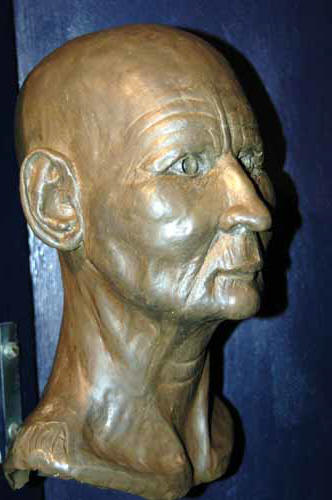 Reconstructed head of Asru Reconstructed head of Asru
-
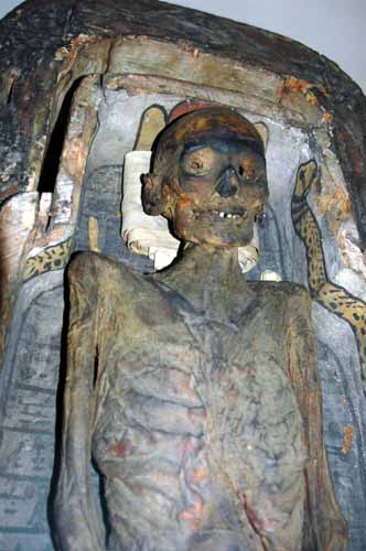
- The mummy of Asru, and the two associated
coffins, were presented to the Manchester museum in 1825 by E. and
W. Garratt. They were the first antiquities of importance in
the Museum's Egyptology collection and they date from c.700 BC. The group probably came from
Thebes (Luxor) in southern Egypt.
-
- The well-preserved body, unwrapped before
it arrived at the Museum, was accompanied by a package of mummified
viscera, placed on the legs. Inscriptions on the coffin provide the
biographical that details that the owner was named Asru and her
mother was Ta-du-Amen and she was a temple chantress. Her duties
involved singing to accompany the sacred rituals to the god Amun.
She may have performed her duties religious centre at the Temple of
Amun at Karnak.
-
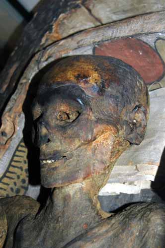
- Mummies are a useful resource for
continuing scientific research and are respected as remains rather than artefacts. The new application of one technique -
immunocytochemistry - has revealed in Asru the presence of the
parasitic disease schistosomiasis. Another technique has enabled us
to identify that aromatic oils were used to anoint her head before
it was bandaged.
-
-
Other
images of Asru
Scientific studies reveal that Asru was probably 50 or 60
when she died. X-rays of her spine show a 'slipped disc' and changes
due to osteoarthritis, as well as abnormal cells in the right
mastoid bone which suggest an earlier infection. These conditions
would have caused chronic back pain and severe earache.-
- Histology has revealed a hydatid cyst in her lung, caused by
a tapeworm Echinococcus Granulosus. As a result of this cyst
Asru would have experienced chest pain and breathlessness.
Electron microscopy has identified a parasite found in part of
her intestine as a nematode worm. Entering the human body
through the skin when the person comes into contact with
contaminated soil, the worm subsequently matures in various
parts of the body and the female lays its eggs in the stomach,
causing severe inflammation. Symptoms would have included pain,
diarrhoea and blood in the faeces. Immunocytochemistry has shown
that Asru also suffered from schistosomiasis. This evidence
occurred in bladder tissue removed from the mummy using an
endoscope. One of the main symptoms would have been blood in the
urine.
The Greater Manchester Police Force devise a procedure to obtain the finger and toe prints and these indicated
that Asru, but the lack of wear, had led a privileged lifestyle.-
- Other studies have included the scientific reconstruction of Asru's
head. Usually, such reconstructions are built up on a cast of the
skull. Here, however, a direct cast cannot be made because it would
damage the tissue on the head. Instead, X-ray computer tomography
has been employed to obtain data of the skull. Using this data, a
sophisticated, numerically controlled milling machine carves a
replica of the skull from a block of polystyrene. The face can then
e built up on this replica skull.
The world's first International Egyptian
Mummy Tissue Bank was set up in the Manchester Museum in 1997. This
provides a new type of museum collection, enabling research to be
carried out on samples obtained from a wide range of mummies
representing various social categories, individual ages, historical
periods and geographical locations within Egypt.
The samples of mummified remains from the
Nile Valley are drawn from collections worldwide (outside Egypt),
and enable investigation of a far larger sample of mummies than
previously possible. Initial studies have focused on
schistosomiasis, but the Bank is available for bona fide research on
other diseases, genetic studies and the investigation of
mummification processes. It has great potential as an international
resource for scientists from a wide range of disciplines. Diagnostic
tools such as immunocytochemistry can be used very effectively to
detect disease in the small samples held in the Bank.
  The International Egyptian Mummy Tissue
Bank at the Manchester Museum is supported with funds from The
Leverhulme Trust and The Kay Hinckley Charitable Trust. Today, the
disease schistosomiasis (Bilharzia) occurs in 74 countries. An
estimated 600 million people are at risk from infection, and 200
million have already contracted the disease. Modern irrigation
schemes and dams have provided new breeding places for the snails
that act as intermediate hosts in the life cycle of the parasite - a
flatworm or blood fluke known as a schistosome. Its eggs cause the
disease. Eggs laid inside the human host are usually discharged into
the water. Some, however, are retained and trigger immunological
responses in the liver, gut wall and bladder. The International Egyptian Mummy Tissue
Bank at the Manchester Museum is supported with funds from The
Leverhulme Trust and The Kay Hinckley Charitable Trust. Today, the
disease schistosomiasis (Bilharzia) occurs in 74 countries. An
estimated 600 million people are at risk from infection, and 200
million have already contracted the disease. Modern irrigation
schemes and dams have provided new breeding places for the snails
that act as intermediate hosts in the life cycle of the parasite - a
flatworm or blood fluke known as a schistosome. Its eggs cause the
disease. Eggs laid inside the human host are usually discharged into
the water. Some, however, are retained and trigger immunological
responses in the liver, gut wall and bladder.
Two species of the parasite - Schistosoma haematobium and Schistosoma
mansoni - cause disease in modern Egypt. Both were present in
ancient times. Today, schistosomiasis is controlled by programmes to
clear snails from the canals, public health education, and mass
chemotherapy using the drug Praziquantel. International teams are
trying to develop a vaccine against the disease. |